Abstract:
In this experiment, we observed and identified red and white blood cells under a light microscope using prepared slides and diagrams. The experiment involved a detailed procedure of sample preparation, observation, and analysis to understand the morphology and characteristics of these blood cells.
Introduction:
The experiment aimed to familiarize students with the identification of red blood cells (erythrocytes) and white blood cells (leukocytes) under a light microscope. Red blood cells are primarily responsible for oxygen transport, while white blood cells are involved in the body's immune response. Observing these cells under a microscope provides insights into their structure, function, and abnormalities that might indicate certain health conditions.
Experiment Details:
The experiment comprised several key steps:
1. Sample Preparation:
We obtained prepared slides containing blood samples. These samples were appropriately stained to enhance the visibility of the cells under the microscope.
2. Microscopic Observation:
We observed the prepared slides under a light microscope, starting with low magnification and gradually increasing to high magnification to visualize the details of individual blood cells.
3. Identification:
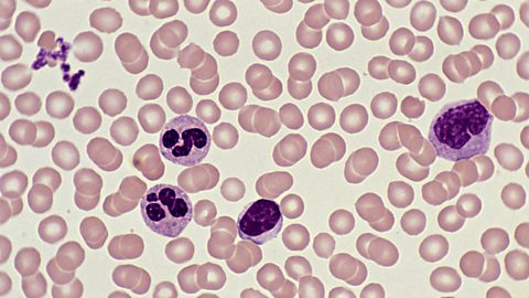
We identified red blood cells based on their characteristic biconcave shape and lack of a nucleus. White blood cells were identified by their larger size, irregular shape, and presence of a nucleus.
4. Morphological Analysis:
We analyzed the morphology of red blood cells, noting variations in size and shape, as well as any abnormalities such as sickle cell shape. For white blood cells, we identified different types (e.g., neutrophils, lymphocytes, monocytes) based on their specific characteristics.
Procedure:
- Obtain prepared slides of blood samples.
- Place the slide on the stage of the light microscope.
- Start observation at low magnification (e.g., 10x objective lens).
- Gradually increase magnification to higher levels (e.g., 40x, 100x) for detailed observation.
- Identify and describe the morphology of red blood cells and white blood cells.
- Record observations and any abnormalities observed.
Observations and Calculations:
Observations:
- Red blood cells: Uniform, biconcave shape, approximately 6-8 μm in diameter.
- White blood cells: Larger, irregular shape, presence of nuclei, various types identified (neutrophils, lymphocytes, monocytes).
Conclusion:
The experiment successfully demonstrated the identification of red and white blood cells under a light microscope. Understanding the morphology and characteristics of these cells is crucial for diagnosing various health conditions and disorders.
Precautions:
- Handle slides and microscope carefully to avoid damage.
- Ensure proper focusing and illumination for clear observation.
- Use appropriate safety measures when handling biological samples.
Short Questions with Answers:
-
What is the purpose of this experiment?
The purpose of this experiment is to identify and differentiate red and white blood cells under a light microscope using prepared slides and visual aids like diagrams and photomicrographs.
-
How are red blood cells (RBCs) identified under the light microscope?
RBCs are identified by their characteristic biconcave shape, lack of a nucleus, and their ability to stain pink/red with certain stains such as eosin.
-
Describe the appearance of white blood cells (WBCs) under the light microscope.
WBCs are larger than RBCs and can have varying shapes. They typically have a nucleus and can exhibit granular or non-granular cytoplasm.
-
What staining techniques are commonly used to visualize blood cells?
Common staining techniques include Wright's stain, Giemsa stain, and Hematoxylin and Eosin (H&E) stain.
-
How can you distinguish between RBCs and WBCs in a prepared slide?
RBCs are smaller, lack a nucleus, and appear pink/red, while WBCs are larger, have a nucleus, and may exhibit various staining properties depending on the type of WBC.
-
What is the function of red blood cells in the body?
RBCs are primarily responsible for transporting oxygen from the lungs to the body tissues and carbon dioxide from the tissues to the lungs for exhalation.
-
Discuss the importance of white blood cells in the immune system.
WBCs play a crucial role in the body's defense against pathogens and foreign invaders. They are involved in phagocytosis, antibody production, and immune response regulation.
-
How can you differentiate between different types of white blood cells?
White blood cells can be differentiated based on their size, shape, staining properties, and the presence or absence of granules in their cytoplasm.
-
Explain the significance of using diagrams and photomicrographs in this experiment.
Diagrams and photomicrographs provide visual aids that help students understand the morphology and characteristics of blood cells more effectively, enhancing the learning experience.
-
What precautions should be taken while handling prepared slides?
Precautions such as proper handling of slides to prevent breakage, avoiding contamination, and using appropriate protective gear like gloves should be taken to ensure safety.
-
Discuss the role of immersion oil in observing blood cells under the microscope.
Immersion oil is used to increase the numerical aperture of the microscope objective, allowing for higher resolution and better visualization of cellular details, including blood cells.
-
How do you calculate the total magnification when observing blood cells?
Total magnification is calculated by multiplying the magnification of the objective lens by the magnification of the eyepiece (ocular) lens.
-
Explain the procedure for preparing a blood smear for observation under the microscope.
A blood smear is prepared by placing a drop of blood on one end of a slide, spreading it thinly using another slide at a 30-45 degree angle, and allowing it to air dry before staining and observation.
-
What are the potential sources of error in this experiment?
Potential sources of error include improper staining techniques, inadequate sample preparation, misidentification of cells, and equipment malfunction.
-
Discuss the differences between bright-field and phase-contrast microscopy in observing blood cells.
Bright-field microscopy uses visible light to observe stained specimens, while phase-contrast microscopy enhances the contrast of unstained specimens by exploiting differences in refractive index.
-
How do you identify abnormal blood cells?
Abnormal blood cells may exhibit irregular shapes, sizes, or staining patterns compared to normal cells. They may also indicate underlying health conditions when observed in a blood sample.
-
Explain the role of a differential white blood cell count in diagnosing medical conditions.
A differential white blood cell count quantifies the percentage of different types of WBCs in a blood sample, which can aid in diagnosing infections, inflammation, and hematological disorders.
-
What are the advantages of digital microscopy in studying blood cells?
Digital microscopy allows for easier storage and sharing of images, precise measurements, and the ability to analyze images using software tools for enhanced research and education.
-
Discuss the ethical considerations involved in obtaining blood samples for scientific research.
Ethical considerations include obtaining informed consent from donors, ensuring the safety and welfare of donors, and respecting privacy and confidentiality in handling personal health information.
-
How can the knowledge gained from this experiment be applied in medical practice?
Understanding blood cell morphology and characteristics is essential for diagnosing and monitoring various medical conditions such as anemia, infections, and leukemia.
Multiple Choice Questions (MCQs) with Answers:
-
What is the main staining technique commonly used to distinguish red and white blood cells under a light microscope?
- Giemsa stain
- Gram stain
- Hematoxylin and eosin stain
- Wright's stain
Correct Answer: a. Giemsa stain
-
Which type of blood cell stains blue-purple and appears larger under the light microscope?
- Red blood cell
- White blood cell
- Platelet
- Neutrophil
Correct Answer: b. White blood cell
-
What is the approximate diameter range of a typical human red blood cell?
- 5-10 micrometers
- 10-15 micrometers
- 15-20 micrometers
- 20-25 micrometers
Correct Answer: a. 5-10 micrometers
-
Which of the following white blood cells is characterized by a lobed nucleus and granular cytoplasm?
- Lymphocyte
- Monocyte
- Basophil
- Neutrophil
Correct Answer: d. Neutrophil
-
When viewing a prepared blood smear under a light microscope, what is the purpose of using the oil immersion technique?
- To increase contrast
- To increase magnification
- To reduce glare
- To improve resolution
Correct Answer: d. To improve resolution
🔗 Other Useful Links
- News By Amurchem
- Free Web Development Course
- All-in-One Exam Prep Portal
- Articles by Amurchem
- Grade 12 Section
- Grade 11 Section
- Grade 10 Section
- Grade 09 Section
- Advanced Artificial Course
- Home and Online Tuition
- Labs By Amurchem
- Science Lectures By Amurchem
- Social Media Executive Course
© 2025 AmurChem. All rights reserved.


