Abstract:
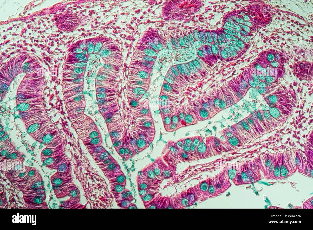
This experiment aims to examine the structure of villi in the small intestine through microscopic analysis of a transverse section. Villi are crucial for absorption in the small intestine, and understanding their morphology is essential for comprehending digestive processes.
Introduction:
The small intestine is a vital organ for digestion and absorption in the human body. Villi, finger-like projections in the small intestine, play a pivotal role in increasing the surface area for absorption of nutrients. This experiment involves the microscopic examination of transverse sections of the small intestine to observe the structure of villi in detail. Understanding the morphology of villi aids in understanding the efficiency of nutrient absorption and various digestive disorders.
Experiment Details:
The experiment involves obtaining a small intestine sample, preparing transverse sections, staining, and observing under a microscope.
Procedure:
- Obtain a small intestine sample from a suitable source.
- Fix the sample in formalin for preservation.
- Embed the sample in paraffin wax for sectioning.
- Prepare transverse sections of appropriate thickness using a microtome.
- Stain the sections with hematoxylin and eosin for better visualization.
- Mount the stained sections on microscope slides.
- Observe the sections under a light microscope, starting with low magnification and gradually increasing to high magnification.
- Record observations of villi morphology, including length, width, and presence of structures like lacteals and goblet cells.
Observations and Calculations:
Observations should include measurements of villi length, width, and presence of structures like lacteals and goblet cells. Formulas for calculating surface area of villi and density of structures can be applied.
Conclusion:
The microscopic examination of transverse sections of the small intestine revealed the intricate structure of villi, which are essential for efficient absorption of nutrients. Understanding villi morphology aids in comprehending the functionality of the small intestine and various digestive disorders affecting nutrient absorption.
Precautions:
- Handle the small intestine sample with care to avoid damage.
- Ensure proper fixation and embedding of the sample for optimal sectioning.
- Use appropriate safety measures when handling chemicals and microscopic equipment.
Short Questions with Answers:
- What are villi, and what is their function?
- Which staining technique is commonly used to visualize tissue structures in microscopic examination?
- What is the purpose of fixing the tissue sample in formalin?
- What is the role of lacteals in the villi?
- How does the structure of villi aid in nutrient absorption?
Villi are finger-like projections in the small intestine that increase the surface area for nutrient absorption. Their function is to enhance absorption efficiency.
Hematoxylin and eosin staining is commonly used for microscopic examination of tissue structures.
Formalin fixation preserves tissue structure by preventing decay and degradation of proteins.
Lacteals are lymphatic vessels in the villi responsible for absorbing dietary fats and fat-soluble vitamins.
The large surface area of villi, combined with microvilli on epithelial cells, facilitates efficient absorption of nutrients by increasing contact between nutrients and absorptive cells.
Multiple Choice Questions:
- Which structure increases the surface area for absorption in the small intestine?
- Liver
- Villi
- Appendix
- Rectum
Answer: b) Villi
- What is the function of goblet cells in the small intestine villi?
- Produce digestive enzymes
- Secrete mucus for lubrication
- Facilitate nutrient absorption
- Initiate peristalsis
Answer: b) Secrete mucus for lubrication
- Which staining technique is commonly used for better visualization of tissue structures in microscopic examination?
- Gram staining
- Hematoxylin and eosin staining
- Methylene blue staining
- Periodic acid-Schiff staining
Answer: b) Hematoxylin and eosin staining
- What is the function of microvilli on epithelial cells of the small intestine?
- Secrete digestive enzymes
- Increase surface area for absorption
- Transport absorbed nutrients to bloodstream
- Produce mucus for protection
Answer: b) Increase surface area for absorption
- Which of the following is responsible for absorbing dietary fats in the small intestine?
- Lacteals
- Goblet cells
- Microvilli
- Paneth cells
Answer: a) Lacteals

